
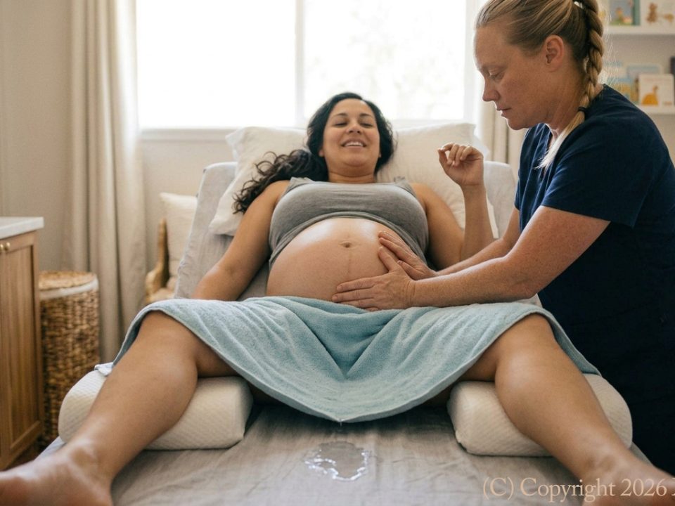
Physiology & Clinical Protocols of Labor Induction Massage
Introduction & Clinical Prerequisites Labor induction massage—more accurately termed labor facilitation massage—operates as a complementary, non-invasive manual therapy designed to optimize maternal biomechanics and autonomic tone prior to labor onset. Crucially, this modality functions strictly to facilitate physiological readiness; it possesses no direct mechanical power to force uterine contractions in an unprepared physiological environment. However, as it’s the present social convention even within medical circles to refer to the modality as Labor Induction Massage Therapy, we shall refer to the practice of facilitation of labor via bodywork and hands-on techniques in this manner, at times.
In clinical practice, manual facilitation requires strict interprofessional collaboration. Therapy must only be initiated following explicit patient consent and a direct prescription or formal clinical clearance from the patient’s primary Obstetrician-Gynecologist. Furthermore, while optimizing neuroendocrine pathways can enhance cervical ripening and contractile coordination, manual therapy cannot resolve structurally or pathologically driven delivery complications. Intrapartum challenges such as cephalopelvic disproportion, placental abruption, or acute fetal distress operate independently of manual intervention and continue to necessitate surgical delivery via Cesarean section regardless of prior facilitation.

Neuroendocrine & Biochemical Pathways
The physiological mechanism of labor facilitation manual therapy centers on downregulating elevated sympathetic tone while activating parasympathetic pathways to trigger specific neuroendocrine cascades. Rather than introducing exogenous pharmaceuticals, this approach optimizes the body’s intrinsic biochemical environment:
Endogenous Oxytocin Secretion & Receptors: Targeted manipulation of cutaneous and deep mechanoreceptors during Labor Induction Massageactivates vagal afferent neural pathways. These sensory impulses travel to the central nervous system, specifically stimulating the paraventricular and supraoptic nuclei of the hypothalamus. This neurogenic reflex promotes the pulsatile release of endogenous oxytocin from the posterior pituitary into systemic circulation. Simultaneously, late-gestational upregulation of myometrial oxytocin receptors (OXTR) causes the uterine smooth muscle to become markedly more responsive to these natural, rhythmically released pulses, facilitating coordinated baseline contractile tone.
Prostaglandin Synthesis & Structural Cervical Ripening: Sustained, focused manual pressure applied along the sacral and pelvic soft tissue structures induces localized hyperemic responses and micro-vascular signaling. This mechanical transduction stimulates uterine, cervical, and fetal membrane cells to synthesize endogenous prostaglandins—specifically prostaglandin E2 (PGE2) and prostaglandin F2α (PGF2α). These local biochemical mediators activate matrix metalloproteinases, which drive extensive extracellular matrix remodeling. This process degrades cervical collagen bundles, increases glycosaminoglycan concentration, and facilitates water absorption, resulting in physical cervical softening, thinning (effacement), and anterior rotation.
Sympathetic Downregulation & Adrenergic Modulation: Chronic maternal stress and anxiety trigger a dominant sympathetic nervous system response, leading to elevated circulating catecholamines (epinephrine and norepinephrine) and cortisol. Epinephrine binds to β2-adrenergic receptors on myometrial cells, directly inhibiting smooth muscle contractions and delaying labor onset. Targeted manual therapy downregulates sympathetic overdrive, causing a marked reduction in systemic cortisol and catecholamines. Removing this β2-adrenergic blockade relaxes the vascular bed, increases uteroplacental perfusion, and restores normal myometrial sensitivity to circulating oxytocin and prostaglandins.
Endorphin Modulation & Ferguson’s Reflex Optimization: By dampening systemic nociceptive signaling through cutaneous afferent stimulation (gate-control theory), manual therapy triggers the central release of endogenous β-endorphins. This natural analgesia prevents the hyper-activation of the maternal hypothalamic-pituitary-adrenal (HPA) axis under stress. As soft tissue resistance is alleviated, fetal presentation can descend and apply uniform mechanical pressure against the ripening internal cervical os. This focal mechanical distortion triggers the neurogenic Ferguson reflex, sending positive feedback signals through pelvic afferents back to the hypothalamus to sustain continuous, self-amplifying oxytocin secretion.

Anatomical Targets & Biomechanical Mechanisms
Manual execution in labor facilitation focuses on key neuromuscular junctions, deep myofascial chains, and somatic reflex vectors to optimize intrapartum pelvic ring compliance and uteroplacental blood flow. The primary anatomical mechanism involves manual modulation of the sacral plexus, specifically targeting spinal segments S2–S4. Applying sustained, non-thrust compressive force across the posterior sacral foramina regulates parasympathetic motor outflow to the pelvic viscera while dampening hyperactivity within the sympathetic hypogastric plexus. This shift in autonomic tone alleviates compensatory myofascial guarding in the piriformis, obturator internus, and levator ani complex. Unwinding these hypertonic pelvic stabilizers reduces mechanical constriction around the internal iliac artery system, enhancing uterine arterial perfusion, optimizing myometrial oxygenation, and establishing the physiological foundation for synchronized, rhythmically efficient contractile signaling.
Complementing this direct neuromuscular modulation is the systematic manipulation of targeted somatic acupressure reflex zones long recognized for their capacity to influence uterine tone and axial alignment. The application of ischemic compression to Sanyinjiao (SP6)—located approximately three cun superior to the prominence of the medial malleolus along the posterior border of the tibia—stimulates afferent neural pathways that cross-communicate with pelvic autonomic ganglia, modulating localized nociceptive signaling and encouraging uterine contractility. Simultaneously, sustained pressure applied to Hegu (LI4), situated deep within the first dorsal interosseous muscle between the first and second metacarpals, engages central descending pain-inhibition pathways. This triggers systemic endorphin release and modifies baseline contractile dynamics without inducing tetanic spasm. To further facilitate fetal descent, manual traction is integrated at Kunlun (BL60), located in the anatomical depression between the high point of the external malleolus and the calcaneal tendon. Working along the superficial back myofascial line, manual stimulation at BL60 releases chronic fascial tension along the posterior chain, easing sacral restriction and encouraging the fetus to descend into the pelvic inlet.
The structural climax of this manual protocol centers on comprehensive pelvic myofascial release, directly targeting the dense connective tissues of the sacrotuberous and sacrospinous ligaments, alongside the muscular sling of the pelvic diaphragm. In late-stage gestation, asymmetric torque or hypertonicity within these ligamentous tethering points can distort the alignment of the sacrum relative to the ilia, restricting the functional diameter of the pelvic canal. Deep, sustained myofascial decompression restores neutral spatial orientation to the pelvic ring, resolving subtle intra-articular torsions. Easing these soft tissue constraints allows the presenting fetal part to achieve ideal occiput-anterior engagement against the pelvic brim. Proper structural alignment ensures that as the fetus descends, it exerts uniform, symmetrical pressure directly against the ripening internal cervical os—a mechanical prerequisite that triggers neurogenic Ferguson reflexes, driving the self-amplifying neuroendocrine feedback loop necessary for spontaneous labor progression.
Safety Profiles, Contraindications, & Risk Mitigation Protocols
Given the direct physiological impact of manual labor facilitation on maternal-fetal hemodynamics and myometrial reactivity, rigorous clinical screening and strict adherence to safety protocols are paramount. While non-pharmacological, manual facilitation exerts powerful systemic effects; consequently, misapplication in high-risk pregnancies presents severe clinical risks, including acute fetal hypoxia, maternal hemorrhage, or emergent uterine rupture.
Absolute Contraindications Manual facilitation is strictly prohibited in the presence of any of the following clinical pathologies:
- Placental & Vascular Pathologies: Placenta previa or low-lying placenta (where manual modulation can trigger life-threatening antepartum hemorrhage), placenta accreta spectrum disorders, and active, unexplained vaginal bleeding.
- Hypertensive & Vascular Disorders: Preeclampsia, eclampsia, chronic hypertension with superimposed preeclampsia, or HELLP syndrome. In these conditions, manual stimulation can unpredictably alter systemic vascular resistance, precipitating acute hypertensive crises or sudden placental abruption.
- Amniotic & Membrane Integrity Compromise: Unmonitored or unconfirmed Premature Rupture of Membranes (PROM) and active chorioamnionitis. Deep tissue manipulation in the setting of ruptured membranes dramatically increases the risk of ascending polymicrobial intrauterine infection.
- Acute Fetal Decompensation & Structural Barriers: Non-reassuring fetal status (e.g., severe recurrent variable decelerations, late decelerations, or sustained fetal tachycardia/bradycardia), transverse or oblique lie, cord presentation or prolapse, and confirmed cephalopelvic disproportion.
Relative Contraindications & Clinical Cautions The presence of relative contraindications demands direct consultation with and written clearance from the attending Obstetrician-Gynecologist prior to initiating therapy:
Prior Uterine Surgery: History of low-transverse Cesarean section or classical uterine incisions (myomectomy), where unmonitored uterine hypertonus could compromise scar integrity.
- Fetal & Gestational Factors: Fetal growth restriction (FGR/IUGR) with oligohydramnios, multiple gestations (twins/triplets), or a history of rapid precipitate labor.
- Maternal Systemic Comorbidities: Insulin-dependent gestational diabetes with vascular complications, active maternal deep vein thrombosis (DVT) or high thromboembolic risk, and severe maternal cardiac disease (NYHA Class II–IV).
Conclusion
Labor facilitation manual therapy represents an integrative, non-invasive approach to birth preparation, bridging neuromuscular manipulation with precise neuroendocrine modulation. By strategically targeting parasympathetic pathways, facilitating endogenous oxytocin and prostaglandin dynamics, and relieving pelvic myofascial restrictions, manual therapy lowers the physiological threshold required for spontaneous labor initiation.
Crucially, this modality functions strictly as an adjunct to optimize baseline maternal mechanics—it cannot mechanically force uterine activity, nor can it resolve acute, underlying structural or pathology-driven obstetric barriers. Because intrapartum emergency events like cephalopelvic disproportion, placental abruption, or fetal distress operate independently of soft tissue manipulation, they remain definitive indications for Cesarean delivery.
When applied within a rigorous safety framework—governed by mandatory Ob-Gyn clearance, thorough screening for absolute contraindications, and vigilant maternal-fetal monitoring—labor facilitation manual therapy serves as a valuable clinical tool to support natural labor onset while upholding the highest standards of maternal and neonatal safety.
